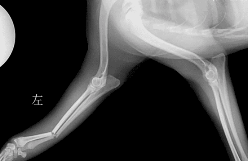
Radial and ulnar fractures are common in small dogs, and they often affect the distal diaphysis. Traumatic fractures of the radius and ulna usually occur mid-shaft, but can occur anywhere. Open traumatic fractures may occur due to limited soft tissue coverage from the mid to distal radius.

Initial diagnosis
Animals with diaphyseal fractures of the radius and ulna are very unstable when first seeking treatment; due to less soft tissue coverage, bone friction sounds are obvious during movement. In order to decide on the best treatment options, it is important to accurately determine the symptoms and medical history.
Age
Skeletally immature animals have good healing abilities, and most dogs under 6 months of age will have a healing time of 6 to 12 weeks if their limbs are sufficiently stable. . However, if misalignment, malunion, and growth plate damage are present, angular deformity of the limb can result as the animal continues to grow. It’s amazing how quickly kittens and puppies less than 10 weeks old heal. Callus can form within 10 days, so if surgical intervention is planned, it should not be delayed.
It takes 2 to 5 months for skeletally mature animals to heal, which will affect how to achieve rigid fixation of fractures, because 5 months of external fixation or external osteosynthesis requires multiple reexaminations, the use of sedatives, Changing bandages, cleaning frames, etc.
Breed
Toy dogs are susceptible to distal radius and ulna fractures. Unfortunately, the rate of nonunion of radius and ulna fractures in toy dogs is high (80% or greater), especially when treated with external osteosynthesis (Figure 1). The poor healing rate is due to a variety of factors, but it is mainly thought to be due to reduced blood vessel density and reduced vascular branching in the distal radius of toy dogs. This means that without rigid fixation of the fracture, usually with plates and screws, these fractures will not heal bonyly.
Craniocaudal and mediolateral radiographs of delayed radius and ulna healing/malunion in a toy poodle cross dog 8 weeks after external splinting. The photo is from the author of this article.
Species
The most important thing to remember is that cats are not small dogs and their anatomy and function are very different, so in cases of radius and ulna fractures cats must be treated carefully Special treatment.
One of the most obvious differences between cats and dogs is that cats’ paws are more prone to supination, which requires some mobility between the radius and ulna. This mobility between bones means that traumatic fractures in cats are more likely to affect the ulna or radius alone than in dogs. This also means that the radius and ulna cannot be internally fixed to each other like in dogs, so each bone will require rigid immobilization if broken.
Cats may have feline patella & dental syndrome (PADS), which may cause non-traumatic fractures of the olecranon process. Therefore, the coexistence of atraumatic fracture and retained deciduous teeth should be evaluated.
Determining Treatment Options
It is easy to focus on a wobbly, broken leg and think that something should be done immediately to fix it. While the next logical course of action may seem to be surgery, before definitive treatment of a fracture it is best to pause and consider whether there may be other injuries that require treatment.

Complicated injuries
Traumatic radius and ulna fractures caused by blunt trauma (such as being hit by a car or falling from a height) may often be associated with complications. Animals should be evaluated for thoracic, abdominal, and neurological injuries.
A thorough clinical examination will help evaluate for the presence of rib fractures, pneumothorax, abdominal wall hernia, and whether the animal is in shock.
A comprehensive neurological examination should be performed, focusing on ruling out head injuries, cervical spine fractures, and brachial plexus injuries.
Horner's syndrome, areflexia, or miosis may indicate brachial plexus injury, and further investigation should be performed if these symptoms are present. Because the radial nerve has the function of innervating the triceps brachii and extensor carpi radialis muscles, the radial nerve is crucial for weight-bearing in the dog's forelimbs.
For many dogs with forearm fractures, retraction and spinal reflexes are difficult to achieve due to pain. Therefore, assessing whether the radial nerve is severely damaged can be accomplished by assessing the superficial sensation on the dorsum of the paw that is innervated by the radial nerve.
While covering the animal's eyes, take a small hemostat and gently pinch the skin on the back of the affected leg's paw. The test is positive if the animal attempts to move its legs away from you and/or signals to stop pinching their paws. This indicates superficial sensation on the dorsum of the paw, and in most cases with normal sensation, complete recovery of radial nerve function occurs within 6 weeks.
Lack of superficial sensation warrants prompt detailed neurologic examination to determine the severity of the injury and prognosis for functional recovery. Be aware, however, that some dogs that are in shock after trauma or after taking large amounts of opioids may show a false negative on this test.
Thorax and abdominal X-rays and ultrasound, as well as an electrocardiogram, can be used to evaluate for rib fractures, pneumothorax and hemothorax, diaphragmatic hernia, myocardial injury (arrhythmia), abdominal wall hernia, urinary tract rupture, and abdominal bleeding.
A comprehensive orthopedic examination is helpful in assessing the presence of any concurrent fractures; however, non-traumatic fractures in cats may require x-rays because the lack of associated soft tissue swelling makes them more difficult to detect. Falls from heights are often associated with ligamentous injuries, so special attention should be paid to assessing the ligamentous support of the wrist and hock joints in these animals.
While examining for concurrent injuries, it may be necessary to place a temporary soft padded bandage ± splint on the fractured forearm to help the animal remain comfortable and to prevent further contamination of the wound.
X-rays of fractures
Craniocaudal and mediolateral radiographs, including those of the elbow and carpus, are usually sufficient to diagnose most radius and ulna fractures. For highly comminuted fractures, oblique radiographs or computed tomography may be helpful to further evaluate whether there is a cleft that may affect treatment options, especially if the cleft is close to the forearm and wrist joint. Direct imaging views are usually easier to obtain without a bandage, but if a wound is present, it should be covered with a dressing and a light bandage to prevent contamination.
Splint, cast or surgery?
The biomechanics of the fracture, mechanical principles, and a variety of clinical factors, including the animal's personality and the client's budget, should be considered when deciding on the final treatment plan. A detailed description of treatment options and their risks and benefits is beyond the scope of this article, but the following three points are worth noting:
1
Any toy dog with radius and ulna fractures should Undergo surgical immobilization, usually rigid internal fixation, and should not be treated with a cast or splint. With the growing popularity of toy poodle crosses, this should also be extended to any breed with a "toy dog characteristic" forearm bone, such as miniature Labradors, Pomskies (a mix of Siberian Huskies and Pomeranians), Italian Greyhound crossbreeds etc. It is important to consider the size of the implant used in these dogs, as an implant that is too large has the potential to cause osteoporosis.
2
Young animals are at risk of limb keratosis after fractures of the radius and ulna. The condition may be worsened if a screw that is too long is used in the proximal radius and a bony syndesmosis develops.
Similarly, if the limb stability is poor, a bony connection and a large amount of callus will form between the radius and ulna, which is most common in external joints.
Although young animals with fractures in both bones may recover quickly, surgical fixation is recommended to minimize the risk of syndesmosis (remember to keep the proximal screws short) rather than fixation with A splint or cast is used to immobilize the leg. However, if only the radius or ulna is fractured in a young animal, the chance of syndesmosis is greatly reduced and external union may be very successful.
3
Cats are not very receptive to forelimb splints or casts (and may even "slough them off" before arriving home). There is also a lot of mobility between the radius and ulna, which is important to protect, but even if just one bone is affected, it can reduce the overall stability of a forearm fracture. Therefore, most forearm fractures in cats can be treated with surgical fixation.
However, very young, healthy kittens have an amazing ability to heal; the author had a six-week-old kitten, and since we had to prioritize some other fractures, we tried to give it A splint was applied instead of surgery; after just two weeks, the fracture had completely healed, and the cat only endured one day of splinting.
Lateral X-ray of a kitten (6 weeks old at the time of fracture) after 3 weeks of resting in a cage, with simple, mild displacement, and complete mid-diaphysis of the radius and ulna. sexual fracture. Both fractures healed without bony union formation or keratinization deformity.
Additional Tips
Young, healthy dogs with only radius or ulna fractures, minimally displaced, closed fractures, require only 6 to 12 weeks of splinting Recovers well with fixation. We prefer to tailor a lateral splint to each case using a plaster bandage (Figure 3) to stabilize the elbow joint (the upper and lower joints must be stabilized if splinting/casting is to be done effectively). Using a plaster bandage eliminates the need for a full cast as we can tailor a strong, form-fitting splint to each animal and use a plaster saw to trim edges etc. as needed.

 扫一扫微信交流
发布评论