1. Basic introduction:
Different types of pigments usually appear in both normal and abnormal liver cytology. Because most of the pigment is within the liver cells and is green when stained with Wright-Giemsa or Diff, special stains are often used to differentiate between different types of pigment.
1. Lipofuscin:
In normal liver cells, most of the pigment is lipofuscin, which is a normal "lossy" pigment that accumulates in lysosomes. of indigestible substances. A lot of granular dark green pigment is seen in the liver cells of older dogs and cats. The accumulation of lipofuscin is considered to be part of the normal aging process and does not directly represent disease. A modified acid-fast dye (Ziehl-Neelsen) was used to confirm the presence or absence of lipofuscin.
2. Bile:
The accumulation of bile in liver cells appears as yellow to oily green or dark green to black pigments of varying sizes. Large amounts of bilirubin, especially when bile-filled duct plugs are present, suggest cholestasis and may be a precursor to clinical jaundice or hyperbilirubinemia. In cytologic specimens, this stasis appears as distinct, intact and fragmented tubular accumulations of dark green, yellow, or olive-green pigment. Hall’s bile stain can be used to confirm the presence of bile (bilirubin). Causes of bile obstruction, such as hepatic lipid deposition or tumor cell infiltration, may be observed in cytology samples, although in many cases the cause cannot be determined.
3. Copper:
Using Diff or Wright-Giemsa staining, copper may be seen in the liver, appearing as rough, granular, light blue, and fragments. like pigments, when present in large amounts, although confirmation requires special staining, such as red acid. Excess copper may cause liver damage or necrosis, accompanied by inflammation. Copper accumulation may be the result of a primary disease, such as a hereditary disease in Bedlington dogs, or a secondary change in chronic hepatitis and other liver diseases.
4. Hemosiderin:
Using Diff or Wright-Giemsa staining, hemosiderin appears as granular yellow-brown to blue-black pigment in the liver cytoplasm. Confirming the diagnosis requires special staining, including Prussian blue staining. Hemosiderin is an iron-containing pigment granule that accumulates in liver cells. It can also appear in disease states, such as hemolytic anemia, chronic inflammation, and when sick animals are subjected to repeated blood perfusion or iron injection.
2. Cytology:

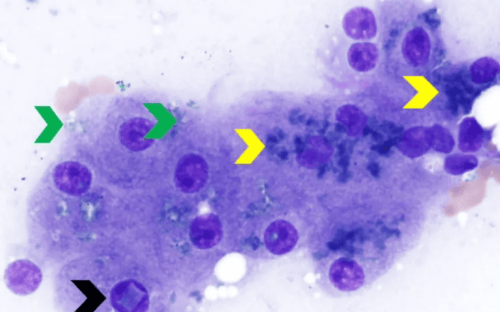
Canine Liver Cytology, Diff 1000X: Note that the granular black-green pigment in the liver cells is lipofuscin (yellow arrow); the black arrow points to the rectangular inclusions in the nucleus, which are occasional and of unknown clinical significance; a small amount of light blue and slightly fragmented in the liver cells The pigment, confirmed to be copper by red acid staining (green arrow).
Feline liver cytology, Wright-Giemsa 1000X: The large amount of granular dark green pigment pointed by the yellow arrow is lipofuscin and comes from clinically normal elderly cats.

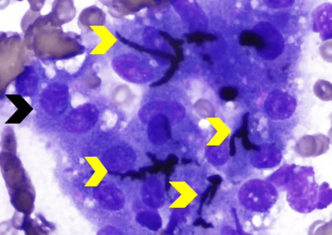
Canine Liver Cholestasis Cytology, Giemsa 1000X: Note clusters of hepatocytes ( Black arrows) are interspersed with plugs of small tubules (yellow arrows) filled with dark green to black bile.
The same canine liver cholestasis cytology, Diff 500X: The yellow arrows confirm that they are several bile-filled tubule plugs, ranging from yellow to oily green.

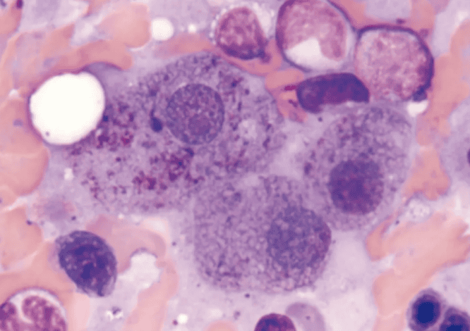
Liver cytology of dogs with chronic hemolytic anemia, Wright's stain 1000X: hemosiderin The pigment appears as a brown-yellow pigment in the liver cells, and the surrounding erythroid and myeloid precursor cells indicate the existence of extramedullary hematopoiesis in the liver.

 扫一扫微信交流
发布评论